
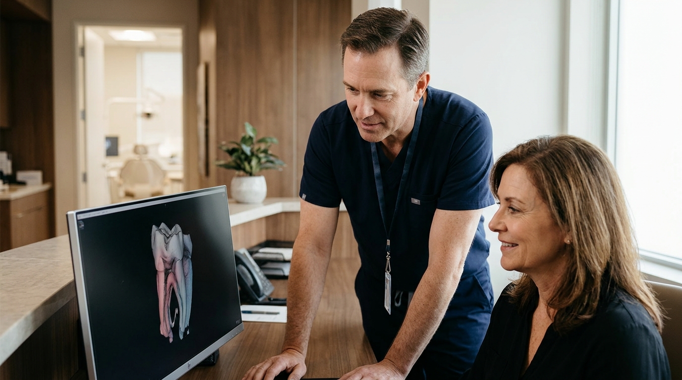
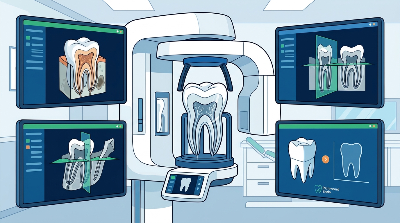
Three-dimensional imaging, specifically Cone Beam Computed Tomography (CBCT), has transformed endodontic care by providing a level of diagnostic clarity that traditional two-dimensional X-rays cannot match. At Richmond Endodontics, we utilize PreXion CBCT imaging to visualize teeth and surrounding structures with unparalleled clarity. This technology allows us to locate all root canals and detect additional pathology, leading to better diagnosis and treatment planning for patients in Eastern Indiana and Western Ohio.
1. Precise Visualization of Complex Root Anatomy
3D imaging reveals hidden canals and complex root shapes that 2D X-rays often obscure.
Many teeth, particularly molars and premolars, possess intricate internal structures that are difficult to see on standard radiographs. CBCT imaging provides a three-dimensional view of the tooth, allowing the endodontist to identify the exact number, shape, and curvature of root canals. This precision is critical for ensuring that every canal is thoroughly cleaned and filled, which is the primary factor in long-term treatment success.
2. Accurate Detection of Pathology
Advanced imaging helps identify infections and bone loss that are invisible on traditional films.
Pathology such as periapical lesions, bone defects, or early signs of infection can be subtle on 2D images. With 3D imaging, we can assess the extent of bone involvement and the precise location of the infection source. This detailed view enables us to formulate a treatment plan that addresses the root cause of the problem rather than just the symptoms, improving the likelihood of healing.
3. Enhanced Diagnosis of Cracked Teeth
3D scans assist in locating fractures that cause intermittent pain and are hard to diagnose.
Cracked teeth often present with symptoms that come and go, making them one of the most challenging conditions to diagnose. While no imaging modality is perfect for detecting cracks, CBCT can sometimes reveal vertical root fractures or other structural defects that contribute to pain. At Richmond Endodontics, we use this technology alongside clinical testing to provide a more confident diagnosis for patients suffering from unexplained tooth pain.
4. Reduced Radiation Exposure
Modern CBCT systems deliver high-definition images with significantly lower radiation doses.
Patient safety is a top priority in our practice. The PreXion CBCT system we utilize provides highly detailed 3D imaging with lower radiation exposure compared to traditional CT scans. This technology allows us to obtain the diagnostic information necessary for successful treatment while prioritizing your safety. We adhere to strict guidelines to ensure that imaging is used only when it adds significant value to the diagnosis.
5. Improved Treatment Planning for Retreatment
3D imaging is essential for evaluating previously treated teeth that are failing to heal.
When a tooth that has already undergone root canal therapy continues to cause pain, retreatment may be necessary. 3D imaging allows us to evaluate the quality of the previous work, identify missed canals, or detect new pathology. This comprehensive view helps us determine whether retreatment, surgery, or another approach is the best option for saving the tooth. Understanding the history and current state of the tooth in three dimensions is vital for a successful outcome.
6. Guided Surgical Precision
3D data supports microsurgical procedures by providing a roadmap for the surgeon.
In cases where endodontic surgery, such as an apicoectomy, is required, 3D imaging provides a detailed map of the surgical site. It helps the surgeon understand the relationship between the root tip and surrounding vital structures, such as nerves and blood vessels. This pre-surgical planning enhances precision and safety, contributing to a smoother recovery and better long-term results for the patient.
7. Better Communication with Patients
Visualizing the problem in 3D helps patients understand their diagnosis and treatment options.
Explaining complex dental issues can be difficult when relying solely on 2D images. By showing patients a 3D view of their tooth and the identified problem, we can make the diagnosis more tangible and easier to understand. This transparency builds trust and ensures that patients are fully informed about their treatment plan. At Richmond Endodontics, we believe that clear communication is a key component of compassionate care.
Choosing the Right Imaging for Your Needs
While 3D imaging offers significant advantages, it is not always necessary for every dental case. The decision to use CBCT is based on the complexity of the diagnosis and the specific clinical findings. If you are experiencing persistent tooth pain or have concerns about a previous root canal, our team will evaluate your case to determine if 3D imaging is the appropriate next step. We are committed to using the most advanced technology available to ensure the best possible outcomes for your dental health.
Frequently Asked Questions
Is 3D imaging painful?
No, 3D imaging is a non-invasive procedure. It involves taking a series of X-rays that are combined to create a 3D image. The process is quick and comfortable, similar to a standard dental X-ray.
How much radiation does CBCT use?
CBCT uses a low dose of radiation, significantly less than a medical CT scan. The amount of radiation is comparable to or slightly higher than a standard dental X-ray, depending on the field of view. We always prioritize patient safety and use imaging only when clinically justified.
Can 3D imaging replace a regular dental X-ray?
3D imaging is a specialized tool used for complex diagnostic cases. It does not replace regular dental X-rays, which are used for routine checkups and monitoring. However, when a specific problem requires a detailed view of the tooth's internal structure, 3D imaging provides superior diagnostic information.
Who should consider 3D imaging for a root canal?
Patients with complex root anatomy, persistent pain after previous treatment, or suspected cracked teeth may benefit from 3D imaging. Your endodontist will recommend 3D imaging if it is necessary to make an accurate diagnosis or plan a complex treatment.
Does Richmond Endodontics use 3D imaging?
Yes, Richmond Endodontics utilizes PreXion CBCT imaging to provide detailed 3D views of teeth and surrounding structures. This technology helps us achieve the highest standard of care and ensure the best possible outcomes for our patients.
Next Steps for Your Care
If you are experiencing tooth pain or have questions about your endodontic treatment, we encourage you to schedule a consultation with our team. At Richmond Endodontics, we are dedicated to providing exceptional treatment in a comfortable and compassionate environment. To plan your visit, to learn more about how advanced 3D imaging can help save your natural teeth.