
What 3D Imaging Reveals About Your Tooth That Regular X-Rays Miss
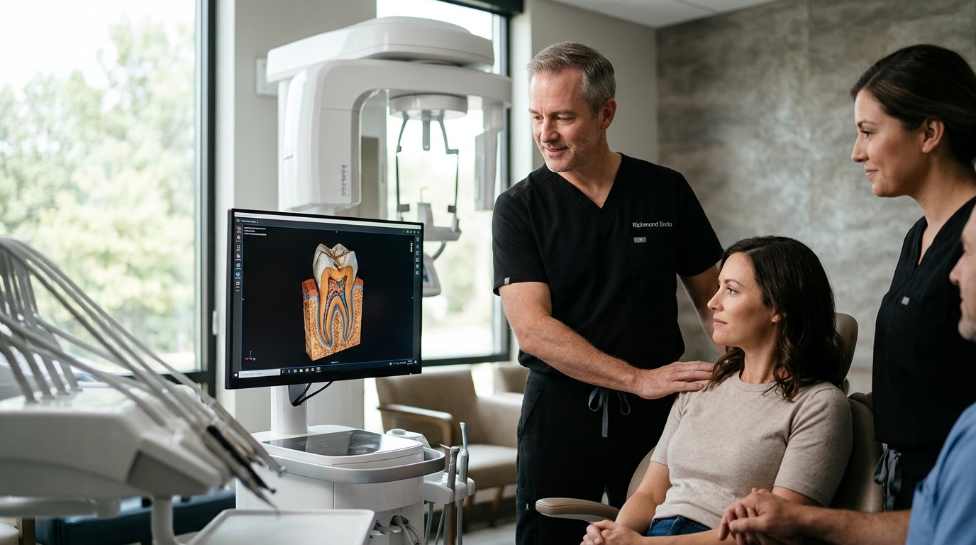
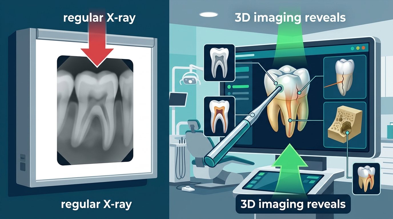
3D imaging reveals the precise location of hidden root canals, fractures, and bone pathology that 2D X-rays flatten and obscure. At Richmond Endodontics, we use PreXion CBCT to visualize your tooth in three dimensions, ensuring accurate diagnosis and treatment planning. This technology allows us to see what is invisible on standard radiographs, leading to better outcomes for complex cases.
The Limitations of Traditional 2D X-Rays
Standard dental X-rays are two-dimensional images of three-dimensional structures. This creates a phenomenon called superimposition, where overlapping tissues can hide critical details. For example, a small crack in a molar or a hidden canal in a premolar may be completely invisible on a 2D film. This limitation can lead to misdiagnosis or incomplete treatment if the clinician relies solely on traditional imaging.
At Richmond Endodontics, we recognize that not every case is straightforward. When a patient presents with persistent pain or a complex anatomical structure, 2D imaging often fails to provide the full picture. We use advanced technology to bridge this gap, ensuring that no detail is overlooked during the diagnostic phase.
How CBCT Provides a 3D Perspective
Cone Beam Computed Tomography (CBCT) is a specialized form of 3D imaging that captures detailed cross-sectional views of the teeth and surrounding bone. Unlike traditional X-rays, CBCT produces a series of slices that can be rotated and viewed from any angle. This allows the endodontist to navigate the root canal system virtually, identifying canals that are curved, calcified, or hidden behind dense bone.
The PreXion CBCT system used at our office provides high-definition 3D images with lower radiation exposure than conventional CT scans. This technology is particularly useful for locating fractures or hidden canals that do not appear on standard X-rays but still manifest pain in the tooth. By visualizing the tooth in 3D, we can plan the most effective treatment strategy with greater confidence.
Clinical Applications of 3D Imaging
3D imaging is essential for several specific clinical scenarios. First, it helps in the diagnosis of cracked teeth, where the fracture line may be too fine to see on a 2D image. Second, it is critical for identifying additional root canals, which are common in molars and can lead to treatment failure if missed. Third, it allows us to assess the proximity of the root tip to vital structures such as the inferior alveolar nerve or the maxillary sinus.
For instance, a patient with a lower molar may have a root that curves sharply toward the nerve. A 2D X-ray might show the root tip near the nerve, but it cannot show the exact distance or the direction of the curve. CBCT provides this spatial relationship, allowing us to perform the procedure safely and effectively. This level of detail is crucial for minimizing risks and maximizing success rates.
Benefits for Patient Comfort and Outcomes
Using 3D imaging leads to more accurate diagnoses and more predictable treatment outcomes. When we can see exactly what is wrong, we can treat it more precisely. This often results in fewer appointments and less discomfort for the patient. Additionally, the ability to plan the treatment in advance reduces the likelihood of complications during the procedure.
At Richmond Endodontics, we are committed to providing the highest standard of care. By utilizing state-of-the-art technology like the PreXion CBCT, we ensure that every patient receives a thorough and accurate diagnosis. This commitment to excellence is reflected in our approach to every case, from simple root canals to complex surgical procedures.
Key Takeaways
- 2D X-rays can miss hidden canals, fractures, and bone pathology due to superimposition.
- CBCT provides a 3D view that allows for precise navigation of the root canal system.
- 3D imaging is critical for diagnosing cracked teeth and assessing nerve proximity.
- The PreXion CBCT system offers high-definition images with lower radiation exposure.
- Accurate 3D diagnosis leads to more predictable outcomes and fewer complications.
- Richmond Endodontics uses advanced technology to ensure comprehensive care for complex cases.
Frequently Asked Questions
Is CBCT safe for patients?
Yes, CBCT is considered safe. It uses a lower dose of radiation compared to conventional medical CT scans and is comparable to or lower than traditional dental X-rays in many cases. The benefits of accurate diagnosis generally outweigh the minimal radiation risk.
Will I always need a 3D scan?
No, not every patient requires a 3D scan. We use CBCT when the case is complex, such as when there is persistent pain, a suspected crack, or unusual anatomy. For straightforward cases, a standard 2D X-ray may be sufficient.
How does 3D imaging help with cracked teeth?
Cracks are often invisible on 2D X-rays. CBCT can sometimes reveal the fracture line or the associated bone changes, helping us determine if the tooth can be saved or if extraction is necessary.
Does 3D imaging reduce treatment time?
While the scan itself takes only a few seconds, the detailed information it provides can streamline the treatment planning process. This can lead to more efficient appointments and fewer unexpected issues during the procedure.
Where can I get 3D imaging in Richmond, Indiana?
Richmond Endodontics offers advanced 3D imaging with the PreXion CBCT system. Our team is experienced in interpreting these images to provide the best possible care for our patients.
Conclusion
3D imaging is a powerful tool that reveals details about your tooth that regular X-rays might miss. By using CBCT, we can diagnose complex conditions more accurately and plan treatments with greater precision. At Richmond Endodontics, we are dedicated to leveraging the latest technology to ensure the best possible outcomes for our patients. If you are experiencing persistent tooth pain or have a complex dental issue, consider scheduling a consultation to see how 3D imaging can help.