
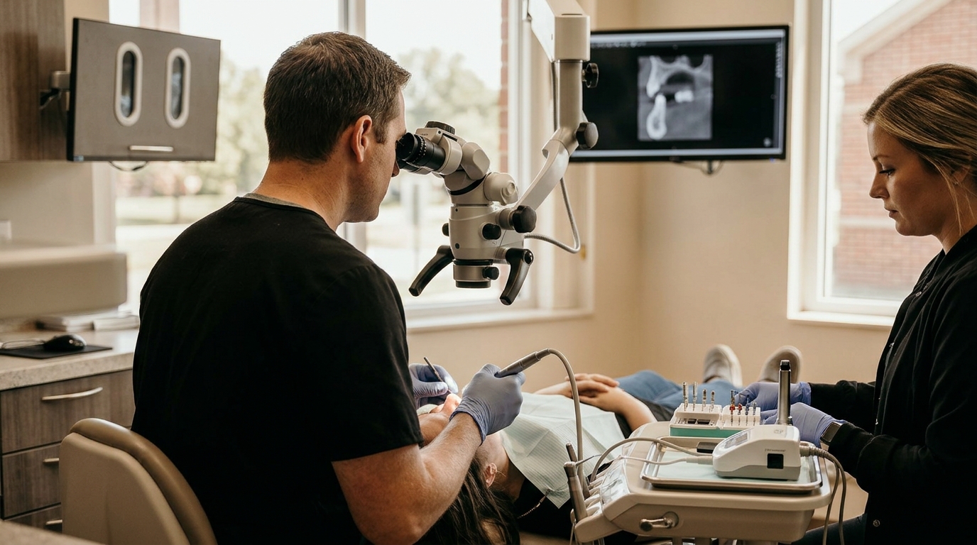
Difficult root canals require specialized tools and expertise to ensure success. Endodontists use advanced imaging, rotary instruments, and microscopic vision to navigate complex anatomy. At Richmond Endo, Dr. Benjamin Adams applies these techniques to treat challenging cases in Eastern Indiana and Western Ohio. This approach improves outcomes for patients with curved roots, calcified canals, or previous failed treatments.
The Role of 3D Imaging in Complex Cases
Traditional 2D X-rays often miss critical anatomical details. Cone Beam Computed Tomography (CBCT) provides a three-dimensional view of the tooth and surrounding bone. This technology allows endodontists to identify hidden canals and fractures that standard imaging cannot show. Accurate diagnosis is the first step in saving a difficult tooth.
Why 3D Matters for Curved Roots
Many molars have highly curved roots that are difficult to clean. 3D imaging helps map the exact curvature and length of these canals. This precision reduces the risk of instrument separation or perforation. Patients benefit from a safer and more predictable treatment plan.
Microscopic Vision and Magnification
Endodontic microscopes provide up to 25x magnification of the tooth interior. This level of detail is impossible to see with the naked eye. The microscope allows the clinician to locate tiny canals and repair perforations. It also helps in removing broken instruments from the root canal system.
Enhancing Precision During Treatment
Working under a microscope changes the dynamic of the procedure. The endodontist can see the texture of the dentin and the flow of irrigants. This visual feedback ensures thorough cleaning and shaping. The result is a cleaner canal that is less likely to harbor bacteria.
Rotary NiTi Instruments and Irrigation
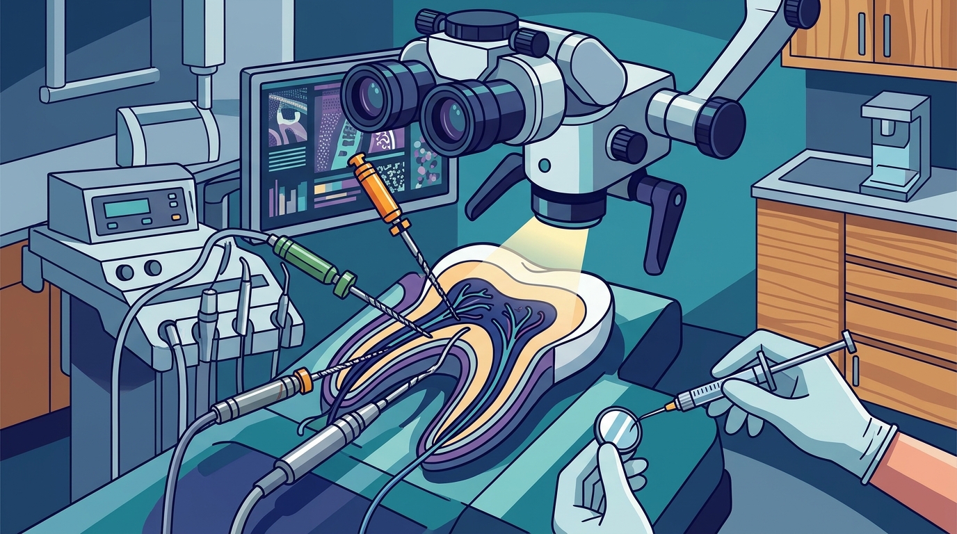
Nickel-titanium (NiTi) rotary files are flexible and resistant to fracture. These instruments can navigate curved canals without binding. Modern files are designed to clean and shape the canal in fewer visits. Efficient instrumentation reduces chair time and patient discomfort.
The Importance of Chemical Irrigation
Mechanical cleaning alone is not enough to disinfect the canal. Sodium hypochlorite and EDTA are used to dissolve organic tissue and smear layer. Ultrasonic activation enhances the penetration of these solutions into complex anatomy. This combination of mechanical and chemical cleaning is critical for long-term success.
Managing Calcified and Obstructed Canals
Calcification occurs when the root canal fills with hard tissue over time. This is common in older teeth or those with previous trauma. Endodontists use specialized burs and piezoelectric tips to remove this obstruction. Patience and precision are required to locate the canal orifice.
When Surgery Is Necessary
If the canal cannot be accessed from the top, an apicoectomy may be needed. This surgical procedure involves removing the root tip and sealing it from the outside. It is a viable option for teeth with persistent infection. Richmond Endo evaluates each case to determine the best non-surgical or surgical approach.
Key Takeaways
- CBCT imaging provides 3D views that detect hidden canals and fractures.
- NiTi rotary files safely navigate curved and complex root anatomy.
- Ultrasonic irrigation enhances the effectiveness of chemical disinfectants.
- Specialized techniques are required to manage calcified or obstructed canals.
- Expertise in both non-surgical and surgical endodontics ensures comprehensive care.
Frequently Asked Questions
What is the most advanced technology used in root canals?
Cone Beam Computed Tomography (CBCT) and dental microscopes are considered the most advanced tools. They provide 3D imaging and high magnification, respectively.
Can a root canal be saved if it was done years ago?
Yes, retreatment is often possible. Advanced techniques allow endodontists to remove old filling material and clean the canal again.
Why are some root canals more difficult than others?
Difficulty depends on root curvature, calcification, or previous trauma. Molars often have multiple canals that are hard to access.
Does using a microscope hurt?
No, the microscope is a tool for the dentist. It improves visibility and precision, which can actually make the procedure more comfortable.
How long does a difficult root canal take?
Complex cases may require two or more visits. The time depends on the specific anatomical challenges of the tooth.
What is an apicoectomy?
An apicoectomy is a surgical procedure that removes the root tip and seals it. It is used when non-surgical treatment is not possible.
Conclusion
Difficult root canals require a combination of technology and skill. Advanced techniques like 3D imaging and microscopy significantly improve success rates. If you are facing a complex dental issue, consult with a specialist. Richmond Endo offers expert care for patients in the region. To schedule a consultation and discuss your treatment options, contact our team. Learn more: Home Richmond Endo.