
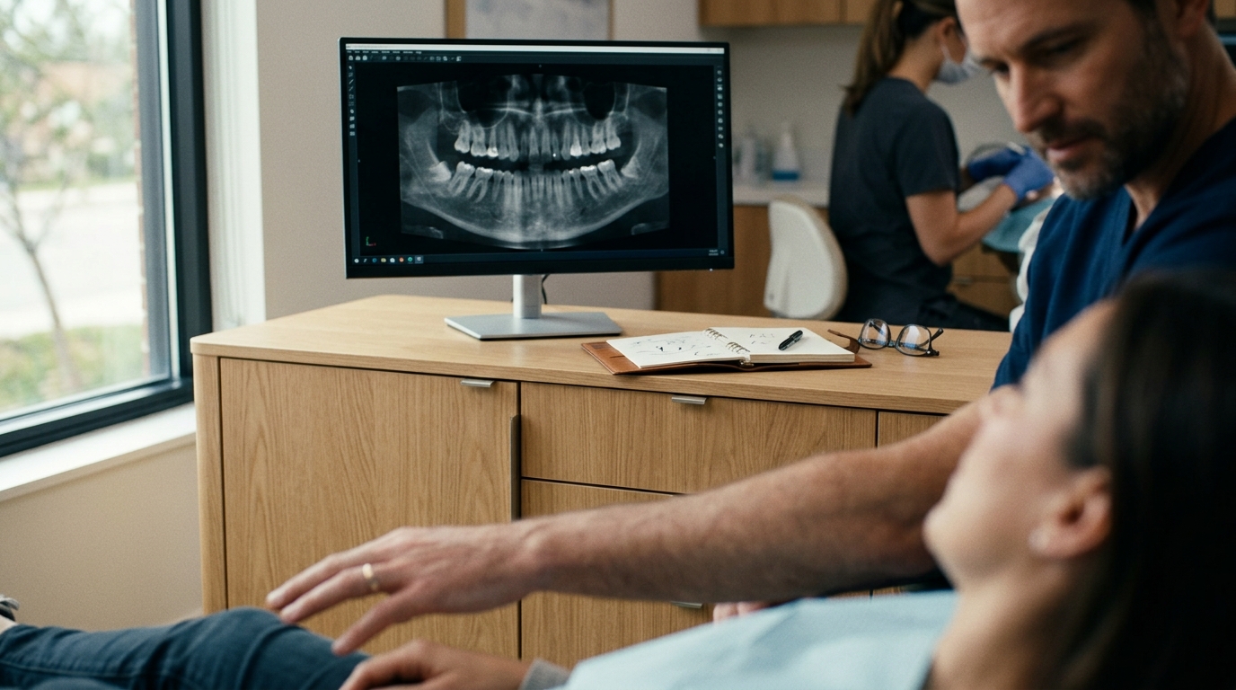
How Endodontists Identify and Diagnose a Cracked Tooth
Endodontists identify cracked teeth by combining detailed patient history, specific biting tests, and advanced imaging to locate fractures invisible to the naked eye. At Richmond Endodontics, Dr. Benjamin Adams utilizes a systematic diagnostic protocol to pinpoint the exact location and depth of the crack. This precise identification determines whether the tooth can be saved through root canal therapy or requires extraction, ensuring you receive the most effective treatment plan for your specific dental condition.
The Clinical Challenge of Cracked Tooth Diagnosis
Diagnosing a cracked tooth is often described as a diagnostic dilemma because the fracture lines are frequently microscopic and do not appear on standard x-rays. A cracked tooth is an incomplete fracture that usually progresses from the crown of the tooth toward the root, often in a mesio-distal direction. Because the crack can close when biting pressure is released, the pain may be intermittent, making it difficult for patients to describe the exact trigger of their discomfort.
Research indicates that despite numerous clinical trials and pre-clinical developments, the diagnosis of cracked teeth, especially in the early stages, remains a significant challenge for dental professionals. The American Association of Endodontists notes that for cracked teeth presenting with irreversible pulpitis, endodontic treatment is a viable option with a favorable prognosis when the crack has not extended below the cementoenamel junction. This distinction is critical because it dictates the long-term survival rate of the tooth.
Physical Examination and Biting Tests
The initial phase of diagnosis involves a thorough physical examination where the endodontist observes the tooth for visible signs of fracture or wear. Dr. Adams performs specific biting tests, such as the Toomey's bite test, to reproduce the patient's pain. When a patient bites down on a specific object, the crack opens, irritating the pulp tissue. When the pressure is released, the crack snaps shut, resulting in a sharp, sudden pain. This specific symptom pattern is a hallmark of cracked tooth syndrome.
During this phase, the endodontist also checks for temperature sensitivities. Patients often report pain when drinking hot or cold liquids, which indicates that the pulp is inflamed or infected. By mapping exactly where the pain occurs during these tests, the specialist can narrow down the location of the fracture. This hands-on approach is essential because it provides immediate feedback that imaging alone cannot offer.
Advanced Imaging and Microscopic Evaluation
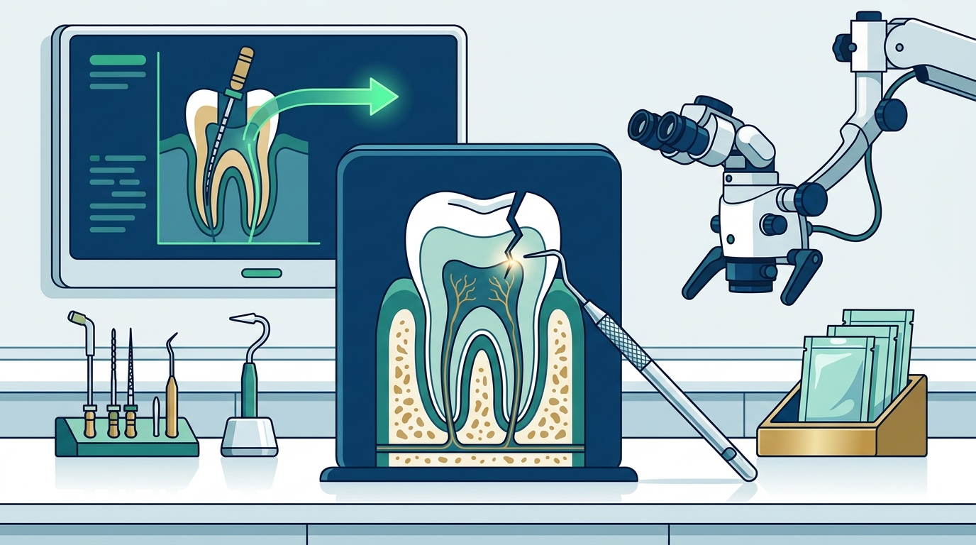
While physical tests are vital, advanced technology provides the definitive evidence needed for a confident diagnosis. At Richmond Endodontics, we utilize the Zeiss ProErgo surgical microscope, which offers superior magnification and lighting. This tool allows our team to detect the finest details in the root canal system that are often invisible to the naked eye. Under high magnification, the endodontist can trace the path of the crack and determine its depth relative to the gum line.
We also employ PreXion CBCT imaging, which provides highly detailed 3D images of the tooth and surrounding structures. Unlike traditional 2D x-rays, CBCT scans can reveal hidden canals and fractures that do not appear on standard radiographs. This 3D visualization ensures we locate all root canals and detect any additional pathology, leading to better diagnosis and treatment planning. The combination of microscopic visual inspection and 3D imaging creates a comprehensive diagnostic picture that minimizes the risk of misdiagnosis.
Interpreting Diagnostic Findings for Treatment Planning
Once the crack is located, the endodontist interprets the findings to determine the appropriate course of action. If the crack is confined to the crown and has not reached the root, the tooth may be saved with a crown or a post and core. However, if the fracture extends below the gum line, the prognosis becomes more complex. In these cases, endodontic surgery, such as an apicoectomy, may be recommended to remove the infected portion of the root tip.
Understanding the depth and direction of the crack is the key to predicting the tooth's longevity. A crack that extends deep into the root structure may eventually split the tooth, necessitating extraction. By identifying the crack early and accurately, Dr. Adams can provide a realistic assessment of the tooth's future. This transparency helps patients make informed decisions about their oral health and financial investment in treatment.
Key Takeaways
- Cracked tooth diagnosis is challenging because fractures are often microscopic and invisible on standard x-rays.
- Specific biting tests help reproduce the sharp pain associated with the crack opening and closing.
- The Zeiss ProErgo microscope allows for high-magnification visual inspection of the tooth structure.
- PreXion CBCT imaging provides 3D views that reveal hidden canals and fractures.
- The depth of the crack relative to the gum line determines the long-term prognosis of the tooth.
- Early diagnosis is critical for preserving the natural tooth and avoiding extraction.
Frequently Asked Questions
Can a cracked tooth be seen on a regular x-ray?
Usually, no. Standard 2D x-rays often fail to show cracks because the fracture lines are too thin and may be obscured by the surrounding bone. Advanced 3D imaging, such as CBCT, is often required to visualize the fracture clearly.
What are the most common symptoms of a cracked tooth?
The most common symptoms include sharp pain when biting down or releasing pressure, sensitivity to hot or cold temperatures, and pain that comes and goes. The pain is often localized to a specific spot on the tooth.
How long does a cracked tooth diagnosis take?
A comprehensive diagnosis typically takes one appointment. The endodontist will perform a physical exam, take advanced imaging, and discuss the findings with you. In complex cases, a second visit may be needed for treatment.
Is a cracked tooth always an emergency?
Not always, but it should be addressed promptly. If the crack extends deep into the root, the tooth may split, leading to infection or the need for extraction. Early intervention offers the best chance of saving the tooth.
Can a cracked tooth be saved?
Many cracked teeth can be saved, depending on the depth and location of the fracture. If the crack is above the gum line, a crown or root canal therapy may be sufficient. If it extends below the gum line, the prognosis is less favorable but still possible in some cases.
Conclusion
Accurate diagnosis is the foundation of successful endodontic treatment. By combining clinical expertise with advanced technology like the Zeiss ProErgo microscope and PreXion CBCT imaging, Richmond Endodontics provides precise diagnoses for complex cases. If you are experiencing pain when biting or sensitivity to temperature, do not ignore the symptoms. Early evaluation by a specialist can save your natural tooth and prevent more invasive procedures in the future.
To schedule a consultation with Dr. Benjamin Adams and our team, please contact our office. We are committed to providing compassionate, expert care for all your endodontic needs in Richmond, Indiana. Learn more: Contact Us Richmond Endo.